Melatonin vs. UV Radiation: A Speculation for Future Therapeutics
Soumik Goswami and Chandana Haldar
Soumik Goswami1 and Chandana Haldar2*
1Visva-Bharati University, Shantiniketan, West Bengal, India
2Department of Zoology, Banaras Hindu University, Varanasi, Uttar Pradesh, India
- Corresponding Author:
- Haldar C
Head, Department of Zoology
Banaras Hindu University
Varanasi- 221005, Uttar Pradesh, India
Tel: +91 9415222261
Fax: +91 542-2575093
E-mail: chaldar2001@yahoo.com
Received Date: September 19, 2016; Accepted Date: October 04, 2016; Published Date: October 07, 2016
Citation: Haldar C, Goswami S (2016) Melatonin vs. UV Radiation: A Speculation for Future Therapeutics. J Clin Mol Endocrinol 1:28. doi: 10.21767/2572-5432.100026
Copyright: © 2016 Goswami S, et al. This is an open access article distributed under the terms of the Creative Commons Attribution License, which permits unrestricted use, distribution, and reproduction in any medium, provided the original author and source are credited.
Abstract
The era of technological boom has brought with it a concealed gift for mankind. However, unlike the literal aspect of “gift”, this has detrimental effects on the living organisms. Being invisible in nature the UV radiations (UVR) continuously bathe the earth without even concerning the organisms which get exposed until off late there has been extensive research on the possible pathologies it inflicts. UVR potentiates several damaging reactions primarily on the skin which in turn affect the whole body homeostasis. Associated with the triggering of oxidative processes the radiation augments the generation of toxic free radicals, the prime initiator of most diseases. An efficient radical scavenger, melatonin proved to be a boon for delineating UVR induced anomalies. Since melatonin is an integral part of the body of almost all living organisms, the possible sideeffects of utilizing commercial products can be avoided by its clinical use. In several studies all over the world involving both in vivo and in vitro approaches, melatonin undoubtedly provided hope for using it as an UVR preventive. The present short review of literature will further summarize the recent advances in melatonin physiology and its use as an anti-UV molecule.
Keywords
Melatonin; UVR; Antioxidant; Apoptosis; Stress; Cellular damage
Environment, UVR and Organisms: The Beginning
Life commenced on this planet after the Big-Bang which was nothing but a series of extensive exothermic reactions comprising toxic gases and other molecules. The effect of sun in carving out the life forms in earth is highly important as it is the sole source of energy. The plants prepare food under photic interventions in the complex process of photosynthesis forming the basal energy level in an ecosystem. The herbivores thrive on the primary producers and followed by the carnivores. Therefore the entire ecosystem sustains itself depending on the solar energy. The shining sun however has its dark side too. Apart from all the life giving attributes the sun is also the only source of UVR. However, the existence of UVR had been eternal but it is only been in recent times that scientists have understood the significance of the damage these causes. One of the main reasons behind this late speculation is the depleting stratospheric ozone layer. Ozone is like a physical barrier for UVRs and it is the vastly spread ozone layer in the stratospheric region that literally protects us from the UVRs. The significant depletion of this ozone layer courtesy the generation and accumulation of chloroflurocarbons (CFCs) has irked the scientific world over the past few decades. The Montreal protocol has been one of the most discussed agenda which vowed to attenuate the production of CFCs and help protect the ozone layer to stop global warming. However, it is not only the warming of the earth which concerns biologists but the ever increasing incidence of UVR penetrance due to the holes in the ozone layer. According to the British Antarctic Survey (BAS) the ozone layer has decreased each September and October since 1977 at a rate of 40% [1]. This is a serious environmental threat to mankind since the entire ozone layer if depleted would spell catastrophical consequences. BAS measurements indicated unexpected ozone depletion in the Antarctic region so severe that computers receiving satellite data often rejected ozone amounts as technical errors [2]. Therefore, the need for protecting the environment is the most significant purpose for the modern world. However, apart from the environmental exposures one can also get exposed to UVRs from electric arc lamps, artificial tanning salons and lighting systems which are becoming a common practice for the modern world [3]. Thus, cumulatively the UVRs induce severe physiological damages due to the rapid interaction with DNA, proteins and triggering the generation of reactive free radicals ultimately culminating in a diseased state.
UVR: The Basics and Biological Attributes
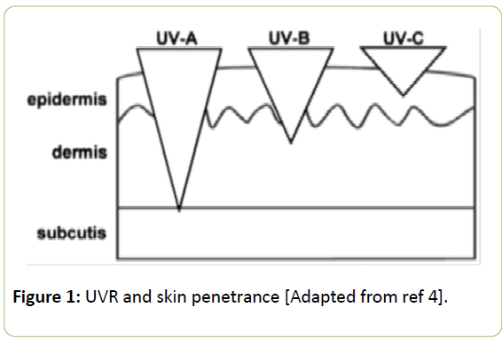
The UVR comprises the wavelengths below the visible spectrum (<400 nm) and that is the reason these are invisible to the naked eye and unlike ionizing radiations are not felt. The entire UV spectrum comprises 3 types of UV rays categorized on the basis of their wavelengths. The lowermost wavelength is attributed to the UVC (<280 nm) while the UVB (280–320 nm) consists of the middle part of the spectrum. The highest wavelength consists of UVA (320–400 nm). The penetrative power of UV depends upon its wavelength and for the skin the UVA has the deepest penetrance followed by UVB and UVC [4]. Altogether all the 3 types of UVRs impart deleterious damage for the body milieu (Figure 1).
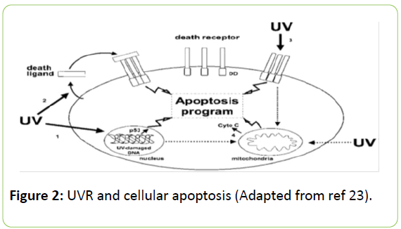
The largest organ of the body is the skin and it is also an innate immune component which forms the very first line of defense against pathogens and other agents perturbing homeostasis. The death of most patients having acute burns is mostly attributed to the disrupted skin barrier and free entry of pathogens. We are invaded by innumerable pathogens and stressors everyday and it is only due to the skin that we do not succumb to them. The UVC rays are mostly blocked in by the ozone and barely reach earth. The matter of concern is the UVA and UVB radiations which readily reach mankind. The UVB penetrates the epidermis completely and reaches dermal layer while the UVA even further reaches to the sub-dermal tissues affecting the innermost components of the skin which comprises immune cells, hormonal machinery and other important glands [4]. However, the skin contains a distinctive defense machinery to counteract the stressors else every single individual would become a sufferer of UVR mediated cutaneous anomalies [5]. The Hypothalamo-Hypophyseal-Adrenal (HPA) axis is the prime system that regulates the stress generated in normal body physiology and acts accordingly to counteract it [6]. The skin has the entire cellular and molecular machinery to synthesize molecules like CRH, POMC, ACTH, β-endorphin and their respective receptors to ameliorate the effects of various damaging agents including UVR [7,8]. A local HPA aids in stabilizing the skin milieu in conditions of severe pathological damages [9]. UVR augments corticotrophin releasing factor (CRF) synthesis locally further inducing the ACTH secretion via the central HPA axis depending on the wavelength of radiation [10]. However, extensive research has shown that these molecular machinery are activated only in case of situations that the local stress management system fail to cater to, as in case of severe UVB irradiation which requires all the anti-stress machinery to unanimously alleviate the perturbation of normal physiology [11]. Oxidative stress refers to shift in the ratio of pro- and antioxidants towards the former and is a normal event of metabolism. One of the most crucial cellular components involved is the mitochondria which comprise the electron transport chain (ETC) and a faulty ETC can lead to electron leakage triggering free radical generation. UVR induces mutagenic issues within the mitochondrial genome termed as “common deletion” which leads to catastrophic electron leakage and active generation of free radicals [12]. Other than mitochondrial damage UVRs also induce oxidation of DNA yielding oxidation products in the form of 8oxo-2dG and cyclobutane pyrimidine dimmers [13]. The wavelength of UV is crucial for the type of cellular damage it inflicts. UVA induces less direct DNA damage on the basis of its incident energy than does UVB [14]. However, UVA is approximately 20 fold more intense in sunlight than UVB and is thus readily absorbed by the skin chromophores [15]. UVB induced DNA mutations are base transitions G:C to A:T [16] while in the case of Chinese hamster ovary cells exposed to UVA, there occur increased transversions i.e., A:T to C:G [17]. The mutagenic effects are associated with immortalization of cells culminating in cancerous growth and UVR is intricately associated with single cell carcinoma and melanoma. The steroids produced within the skin play a protective role against the tumorigenicity by getting converted into secosteroids thereby providing protection [18,19]. The other effect of UVR is the induction of sunburn which is characterized by pyknotic cells having shrunken cytoplasm and membrane blebbing indicating the induction of apoptosis [20]. These apoptotic cells are the dermal fibroblasts which suffer the most under the incidence of UVR. The arrests of the cells at G1 phase of the cell cycle are mediated by p53 and hence there exists a crucial role of p53 in UV mediated cellular apoptosis [21]. UVRs also trigger death receptor mediated apoptotic pathways involving TNFR and CD95L and activation of these molecules following UVC radiation further substantiates the direct role of UV in cellular apoptosis [22]. Similarly single UV exposure has been investigated to cause a decline in bcl-2 transcripts in rat skin [23] thus providing experimental evidences of the great amount of damage UV can cause. Along with the skin the UVR is also associated with affecting systemic physiology. Although the radiation does not reach internal organs the locally generated free radicals in the skin get transported to the internal milieu and put the systemic homeostasis at jeopardy. This has been designated as “bystander effects” of UVR and is out of the scope of this review (Figure 2).
Melatonin: The Molecule of Darkness against a Component of Sun-Light
An accidental discovery by Prof. A. B. Lerner in 1958 the neuro-hormone melatonin was so named due to the blanching effect on the melanophores of tadpole skin [24]. Ever since the discovery of this molecule the world scientific community has always been intrigued by the wide plethora of physiological functions it governs. The pineal gland is the primary source of the indole-amine although gradually melatonin synthesis has been associated with a variety of organs including the skin [25]. The first precursor for the biosynthesis is the amino acid tryptophan and downstream steps of hydroxylation, decarboxylation and methylation yields the final product. Until recently, the enzyme aryl alkylamine N-acetyl transferase (AANAT) was considered to be the rate limiting enzyme for melatonin biosynthesis but endeavors by Slominski et al. have provided sufficient experimental evidences to prove that AANAT is not the rate limiting enzyme for melatonin biosynthesis and in absence of AANAT, arylamine NAT (ANAT) can trigger acetylation of serotonin required for the pathway to continue [26,27]. The most interesting feature of melatonin synthesis is the rhythmicity it follows peaking at night hours and decreasing at day. Hence, the duration of the short-lived molecule in blood signifies the hour of the night. Melatonin primarily exhibits its physiological functions acting via G protein coupled membrane receptors although the existence of cytosolic and nuclear binding sites has also been evidenced [28]. Melatonin regulates significant physiological functions including reproduction, immune functions, apoptosis, tumorigenesis and oxidative load. Among all the different functions the action of melatonin as an antioxidant is the most vital aspect that provided the basis for using it against UVR. Melatonin is an active free radical and a terminal antioxidant with the ability to reach intricate areas of cells owing to its lipophilicity and can even breach the blood brain barrier [29]. The most important aspect of melatonin as an antioxidant is that even the by-products of melatonin metabolism (AFMK, AMK) act as potential radical scavengers. Dietary antioxidants like vitamin C often undergo redox cycling and become pr-oxidative which is not in case of melatonin which reacts with free radicals to form stable end products [30]. Scientific insights into the scavenging property of melatonin has proved that the capacity to scavenge free radicals is much higher than other antioxidants and a single molecule of melatonin can react with 4 reactive species and turn them into harmless products [31]. The most important function of melatonin that places it above all other antioxidants is that apart from neutralizing the radicals, melatonin also up regulates the gene expression of crucial anti-oxidative enzymes like superoxide dismutase (SOD) and glutathione peroxidase (GSH-Px) while in case of catalase the action of melatonin depends on the tissue milieu [32].
Melatonin vs. UVR: A Preventive Approach
“Prevention is better than cure”, an age old proverb proves extremely true while discussing the anti-UVR attributes of melatonin. Thus, it is to be noted that the efficacy of melatonin against UVR depends highly on the time of its administration. Photo-biologists after extensive research have concluded that melatonin is more effective when administered before the onset/irradiance of UVR than when applied after the UVR has been irradiated [33]. This explains that melatonin needs to be in the milieu of UV incidence in a higher concentration than physiological levels to exert its beneficial properties. Melatonin when administered at concentrations of 10-3 and 10-4 M elicited beneficial and protective properties in UV irradiated epidermal keratinocytes [34]. As discussed earlier melatonin is short-lived and is metabolized rapidly. However, the metabolites of melatonin also show protective property against UVR. Reports have shown that UVB irradiation induced significant free radical generation in epidermal keratinocytes which got reverted by the metabolites of melatonin [35,36]. The most recent discovery in the field of cutaneous photo-biology is the existence of an orchestrated defense system depicted as the Melatonergic Anti-oxidative System (MAS) which provides protection from different biotic and abiotic stressors [37]. The MAS is attributed not only to the superficial epidermal tissue but the underlying dermal gland and hair follicles since UVA has deepest penetrative power to affect these sub-dermal structures.
Therefore, the application of melatonin as formulation or supplement would definitely provide significant protection against UVR. The mode of administration of melatonin best suited for ameliorating UVR mediated anomalies is in the form of topical dermal formulations. The low molecular weight and short plasma half-life of this hormone makes it a suitable compound to be incorporated in such formulations against UV radiation [38,39]. The topical application of melatonin builds a depot in the stratum corneum region of the upper epidermis and is gradually released into the dermis and blood vessels where it can exert its biological actions [40]. The anti-apoptotic actions of melatonin also involve the preservation of mitochondrial membrane integrity and thereby inhibiting the UV radiation induced cellular apoptosis [2]. Cumulatively all these extensive studies definitely aim at considering melatonin as a target molecule to counteract the deleterious effects of UVR.
Conclusion
The sole source of light and energy also exhibit damaging effects of great concern for mankind. The extent of urbanization is inevitable and so is the risk of getting exposed to UVRs. Moreover, the practices of artificially getting tanned have also become a significant priority further summing up the risks of greater damaging consequences. The commercial applications do have benefits against the UVR but the physiological effects are still significantly prevalent as statistical data reveals increasing incidences of skin carcinomas. Thus, it is high time that melatonin should be incorporated in dermal formulations owing to its multi-faceted physiological functions in providing protection not only against the oxidative potency of UVR but also against the tumorigenic and inflammatory responses triggered by UVRs. Although further extensive research needs to be performed before prescribing melatonin as a drug against UVR, yet it is the future prognostic molecule which would be of great use to mankind to delineate the detrimental damages induced by UVR.
Acknowledgment
The authors report no conflict of interests personal, financial or professional. The financial assistance to Dr. Soumik. Goswami from UGC-DSK is highly acknowledged. The authors acknowledge the instrument gift from Alexander von Humboldt foundation, Bonn, Germany and BRNS-DAE (2010/37B/19/BRNS) for financial assistance in the form of research grants to Prof. C. Haldar.
References
- Farman JC, Gardiner BG,Shanklin JD (1985)Large losses of total ozone in Antarctica reveal seasonal ClOx/NOx interaction. Nature 315: 207–210.
- Goswami S, Haldar C (2015) Melatonin as a possible antidote to UV radiation induced cutaneous damages and immune-suppression: An overview. J PhotochemPhotobiol B 153: 281-288.
- Schröder P,Krutmann J (2005)Enviromental oxidative stress—environmental sources of ROS. Handb Environ Chem 2: 19–31.
- D'Orazio J, Jarrett S, Amaro-Ortiz A, Scott T (2013) UV radiation and the skin. Int J MolSci 14: 12222-12248.
- Slominski A, Wortsman J (2000) Neuroendocrinology of the skin. Endocr Rev 21: 457-487.
- Chrousos GP (1995) The hypothalamic-pituitary-adrenal axis and immune-mediated inflammation. N Engl J Med 332: 1351-1362.
- Slominski A, Wortsman J, Luger T, Paus R, Solomon S (2000) Corticotropin releasing hormone and proopiomelanocortin involvement in the cutaneous response to stress. Physiol Rev 80: 979–1020.
- Slominski AT, Zmijewski MA, Skobowiat C, Zbytek B, Slominski RM, et al. (2012) Sensing the environment: regulation of local and global homeostasis by the skin's neuroendocrine system. AdvAnatEmbryol Cell Biol 212: 1-115.
- Skobowiat C,Dowdy JC, Sayre RM, Tuckey RC, Slominski A (2011) Cutaneous hypothalamic pituitary–adrenal axis homolog: regulation by ultraviolet radiation. Am J PhysiolEndocrinolMetab 301: E484–E493.
- Jozic I, Stojadinovic O, Kirsner RS, Tomic-Canic M (2015) Skin under the (Spot)-Light: Cross-Talk with the Central Hypothalamic-Pituitary-Adrenal (HPA) Axis. J Invest Dermatol 135: 1469-1471.
- Slominski AT (2015) Ultraviolet radiation (UVR) activates central neuro-endocrine immune system.PhotodermatolPhotoimmunolPhotomed 31: 121–123.
- Berneburg M, Grether-Beck S, Kurten V, Ruzicka T, Briviba K (1999) Singlet oxygen mediates the UVA-induced generation of the photoaging associated mitochondrial common deletion. JBiolChem 274: 15345.
- Zhang X, Rosenstein BS, Wang Y, Lebwohl M, Wei H (1997)Identification of possible reactive oxygen species involved in ultraviolet radiation-induced oxidative DNA damage. Free Rad Biol Med 23: 980–985.
- Setlow RB (1974) The wavelengths in sunlight effective in producing skin cancer: a theoretical analysis. ProcNatlAcadSci USA 71: 3363-3366.
- Agar NS, Halliday GM, Barnetson RC, Ananthaswamy HN, Wheeler M, et al. (2003) The basal layer in human squamous tumors harbors more UVA than UVB fingerprint mutations: a role for UVA in human skin carcinogenesis, ProcNatlAcadSci USA 101: 4954–4959.
- Brash DE, Rudolph JA, Simon JA, Lin A, McKenna GJ, et al. (1991) A role for sunlight in skin cancer: UV-induced p53 mutations in squamous cell carcinoma. ProcNatlAcadSci USA 88: 10124-10128.
- Drobetsky EA, Turcotte J, Chateauneuf A (1995) Mutagenic specificity of solar UV light in nucleotide excision repair-deficient rodent cells. ProcNatlAcadSci USA 92: 2350–2354.
- Slominski AT, Zmijewski MA, Semak I, Zbytek B, Pisarchik A, et al. (2014) Cytochromes p450 and skin cancer: role of local endocrine pathways. Anticancer Agents Med Chem 14: 77-96.
- Slominski AT, Manna PR, Tuckey RC (2014)Cutaneous glucocorticosteroidogenesis: securing local homeostasis and the skin integrity.ExpDermatol 23: 369–374.
- Daniels F, Brophy D, Lobitz WC (1961) Histochemical responses of human skin following ultraviolet irradiation. J InvestigDermatol 37:351–357.
- Ziegler A, Jonason AS, Leffell DJ, Simon JA, Sharma HW, et al. (1994) Sunburn and p53 in the onset of skin cancer. Nature 372: 773-776.
- Leverkus M, Yaar M, Gilchrest BA (1997) Fas/Fas ligand interaction contributes to UV-induced apoptosis in human keratinocytes. Exp Cell Res 232: 255-262.
- Gillardon F, Eschenfelder C, Uhlmann E, Hartschuh W, Zimmermann M (1994) Differential regulation of c-fos, c-jun, jun B, Bcl-2 and Bax expression in rat skin following single or chronic ultraviolet radiation and in vivo modulation by antisense oligodeoxynucleotidesuperfusion. Oncogene 9: 3215–3219.
- AB Lerner, JD Case, Y Takahashi (1958) Isolation of melatonin, a pineal factor that lightens melanocytes. J Am ChemSoc 80: 2057–2058.
- Slominski A, Baker J, Rosano TG, Guisti LW, Ermak G, et al. (1996) Metabolism of serotonin to N-acetylserotonin, melatonin, and 5-methoxytryptamine in hamster skin culture. J BiolChem 271: 12281-12286.
- Slominski A, Pisarchik A, Semak I, Sweatman T, Szczesniewski A, et al. (2002) Serotoninergic system in hamster skin. J Invest Dermatol 119: 934-942.
- Reiter RJ, Tan DX, Terron MP, Flores LJ, Czarnocki Z (2007) Melatonin and its metabolites: New findings regarding their production and their radical scavenging actions. ActaBiochim Pol 54: 1–9.
- Slominski RM, Reiter RJ, Schlabritz-Loutsevitch N, Ostrom RS, Slominski AT (2012) Melatonin membrane receptors in peripheral tissues: distribution and functions.Mol Cell Endocrinol 351: 152–166.
- Pandi-Perumal SR, Srinivasan V, Maestroni GJ, Cardinali DP, Poeggeler B, et al. (2006) Melatonin: Nature's most versatile biological signal? FEBS J 273: 2813-2838.
- Tan DX, Reiter RJ, Manchester LC, Plummer BF, Limson J (2000) Melatonin directly scavenges hydrogen peroxide: a potentially new metabolic pathway of melatonin biotransformation, Free RadicBiol Med 29: 1177–1185.
- Tan DX, Reiter RJ, Manchester LC, Yan MT, El-Sawi M (2002) Chemical and physical properties and potential mechanisms: melatonin as a broad-spectrum antioxidant and free radical scavenger. Curr Top Med Chem 2: 181–198.
- Tomás-Zapico C, Coto-Montes A (2005) A proposed mechanism to explain the stimulatory effect of melatonin on antioxidative enzymes. J Pineal Res 39: 99-104.
- Slominski A, Wortsman J, Tobin DJ (2005) The cutaneous serotoninergic/melatoninergic system: securing a place under the sun. FASEB J 19: 176–194.
- Fischer TW, Zbytek B, Sayre RM, Apostolov EO, Basnakian, et al. (2006)Melatonin increases survival of HaCaT keratinocytes by suppressing UV-induced apoptosis. J Pineal Res 40: 18–26.
- Janjetovic Z, Nahmias ZP, Hanna S, Jarrett SG, Kim TK, et al. (2014) Melatonin and its metabolites ameliorate UVB-induced damages in human epidermal keratinocytes. J Pineal Res 57: 90–102.
- Slominski AT, Kleszczya ski K, Semak I, Janjetovic Z, Zmijewski MA, et al. (2014) Local melatoninergic system as the protector of skin integrity. Int J MolSci 15: 17705-17732.
- Fischer TW, Sweatman TW, Semak I, Sayre RM, Wortsman J, et al. (2006) Constitutive and UV-induced metabolism of melatonin in keratinocytes and cell-free systems. FASEB J 20: 1564-1566.
- Fischer TW, Scholz G, Knoll B, Hipler UC, Elsner (2001) Melatonin reduces UV induced reactive oxygen species in dose-dependent manner in IL-3-stimulated leukocytes. J Pineal Res 31: 39–45.
- Fischer TW, Scholz G, Knoll B, Hipler UC, Elsner P (2004) Melatonin suppresses reactive oxygen species induced by UV irradiation in leukocytes. J Pineal Res 37: 107-112.
- Bangha E, Lauth D, Kistler GS, Elsner P (1997) Daytime serum levels of melatonin after topical application onto the human skin. Skin Pharmacol 10: 298-302.
Open Access Journals
- Aquaculture & Veterinary Science
- Chemistry & Chemical Sciences
- Clinical Sciences
- Engineering
- General Science
- Genetics & Molecular Biology
- Health Care & Nursing
- Immunology & Microbiology
- Materials Science
- Mathematics & Physics
- Medical Sciences
- Neurology & Psychiatry
- Oncology & Cancer Science
- Pharmaceutical Sciences


